Digital x-ray
X-ray imaging (radiography) remains the maximum generally used approach in radiology. To create a radiograph, part of body to be examined is subjected to a small amount of X-rays. The X-rays travels via the tissues, to the detector to create a picture. X-rays are usually safe if done by expert radiologists and skilled technologists with minimal possible exposure. No radiation stays after the radiograph is obtained. X-ray is used to examine a part of the body and are used maximum generally to search for fractures. They also are generally used to have a look at the lungs, abdomen, and superficial tissues. X-rays can pick out many fractures, stones

WHY DIGITAL X-RAYS ARE BETTER THAN TRADITIONAL X-RAYS
Radiography is a treasured diagnostic device which assist physicians in a vast majority cases, from pneumonia and bone fractures to cancerous and noncancerous tumors. But, because of the fantastic advances in healthcare, establishments are shifting from conventional X-rays to digital radiography has been proven to make the complete system simpler, faster, and cost-effective than ever before. In fact, while in comparison to conventional radiography, digital X-rays provide many a kind blessings which result in superior patient care.The Advantages of Digital X-Rays
Of course, conventional radiography isn't obsolete. It has been round for properly over a century and has performed a essential position in lots of clinical advancements. However, digital radiography is proving to be relatively safer and convenient diagnostic pictures.Ultrasound
What an ultrasound is?
An ultrasound is an imaging check that makes use of sound waves to create photograph of organs, tissues, and different systems of the frame. Unlike x-rays, ultrasound don’t use any radiation. It is absolutely safe in pregnancy. An ultrasound also can display components of the body in motion, consisting of a beating heart or flowing blood in the blood vessels.
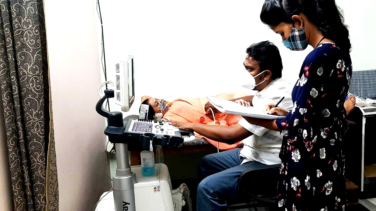
Ultrasound is an most trusted device in antenatal testing and follow up of a pregnant women. In pregnancy it is used to study foetal growth and other parameters. The check can offer facts about baby’s growth, improvement, any abnormality (anomaly scan) and well being of babies.
Diagnostic ultrasound is used to view particular facts about different inner components of the body. These encompass the stomach, thyroid, breasts, cardiac structures, blood vessels, liver, bladder, kidneys, male and female reproductive system anatomy.
Other names: Sonography, ultrasonography, FPP sonography, fetal ultrasound, obstetric ultrasound, diagnostic medical sonography, diagnostic scientific ultrasound
What is it used for?
An ultrasound may be done for prognosis of a disease to development and tracking of a health issue. Growth of fetus inside the uterine cavity.
Pregnancy ultrasound is performed to get data about the wellness of an unborn child. It can be used to:
Confirm whether you are pregnant.
Check the dimensions and position of the unborn child.
Estimate expected date of delivery (EDD). This is referred to as gestational age.
Check for evidence of Down syndrome, which encompass nuchal thickening (of the child's neck).
Check for birth defects of baby's brain, spinal cord, heart, extremities and other body parts.
Doppler ultrasound
Doppler ultrasound is a noninvasive test that may be used to estimate the blood flow inside your blood vessels, with the help of bouncing high-frequency sound waves (ultrasound) of circulating RBS's (red blood cells). A normal ultrasound makes use of sound waves to provide pictures, however can not display blood flow.
A Doppler ultrasound can estimate how rapid blood flows by measuring the rate of alternate in its pitch (frequency). During a Doppler ultrasound, a technician skilled in ultrasound imaging (sonographer) presses a small handheld device (transducer), approximately the dimensions of a fist, on your body surface to be examined, transferring from one place to every other as necessary.
A Doppler ultrasound may also aid in diagnosis of many conditions, including:
Thrombus, clots, malfunctioning valves in your veins, that can cause blood or different fluids to
accumulate abnormally (venous insufficiency)
Heart valve defects and congenital heart disease
A blocked artery (arterial occlusion)
Decreased blood flow into your legs (peripheral artery disease)
Doppler echocardiogram
Best Diagnostic Center which provide best services
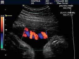
Sound waves change pitch once they bounce off blood cells moving in your heart and blood vessels. These changes (Doppler signals) can assist your doctor measure the rate and path of the blood flow to your heart.
Doppler strategies are typically utilized in transthoracic and transesophageal echocardiograms. Doppler strategies also can be used to test blood flow issues and blood pressure in the arteries of your heart — which conventional ultrasound may not detect.
The blood flow shown on the monitor is colorized to assist your doctor pinpoint any issues.
Risks
No dangers are involved in a transthoracic echocardiogram. You may experience some pain from the transducer being held very firmly towards your chest. The firmness is essential to supply the fine pictures of your heart.
How you prepare?
No arrangements are needed for a standard transthoracic echocardiogram. You can consume, drink and take medicines as you commonly would.
What you may expect?
For a transthoracic echocardiogram:
You'll undress the lower chest and lie on the bed.
Echocardiography
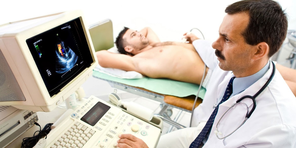
Echocardiography or echo, is a painless test that makes use of sound waves to create moving pics of your heart. The images display the dimensions and form of your heart. They additionally display how nicely your heart's chambers and valves are working. This common test permits your doctor to look at your heart's health.
Depending on what facts your physician desires, you could have one in all numerous forms of echocardiograms. Each kind of echocardiogram entails few, if any, dangers.
Why it is done
Your physician may recommend an echocardiogram to:
Check for issues with the valves or chambers of your heart
Check if heart issues are the reason for signs and symptoms which includes shortness of breath or chest pain
Detect congenital heart defects earlier than birth (fetal echocardiogram)
Transthoracic echocardiogram
In this popular type of echocardiogram:
A technician (sonographer) spreads gel on a device (transducer).
Holter Monitoring
A Holter monitor is a small, wearable device that records a non-stop ECG, generally for twenty-four to forty eight hours.
Event monitor. This transportable device is much like a Holter monitor, however it records mostly at positive instances for a couple of minutes. You can put on it longer than a Holter screen, generally 30 days. You commonly push a button when you experience signs. Some gadgets automatically document when an atypical rhythm is detected.
Event monitor. This transportable device is much like a Holter monitor, however it records mostly at positive instances for a couple of minutes. You can put on it longer than a Holter screen, generally 30 days. You commonly push a button when you experience signs. Some gadgets automatically document when an atypical rhythm is detected.
A Holter monitor is a machine that continuously records the heart's rhythms. The monitor is worn for 24 to 48 hours during normal activity.
Electrodes (small conducting patches) are stuck onto your chest. These are attached by wires to a small recording monitor. You carry the Holter monitor in a pocket or pouch worn around your neck or waist. The monitor runs on batteries.
While you wear the monitor, it records your heart's electrical activity
Keep a diary of what activities you do while wearing the monitor, and how you feel.
After 24 to 48 hours, you will return the monitor to your health care provider's office.
Polyclinic
A polyclinic is a medical facility that is larger than an individual clinic, but smaller than a hospital. It is a medical centre where a patient can meet a number of doctors, get pathological tests and minor procedures done. ... Think of a polyclinic as an upgraded version of a regular medical clinic.
A polyclinic (where poly means "many"; not to be confused with the homonym policlinic, where poli means "city" and which is sometimes used for a hospital's outpatient department) is a clinic or health care facility that provides both general and specialist examinations and treatments for a wide variety of diseases and injuries to outpatients and is usually independent of a hospital.[1][2][3] When a polyclinic is so large that it is in fact a hospital, it is also called a general hospital.[4]
A clinic may be defined as a clinical establishment providing examination, consultation, prescription to outpatients including dispensing of medicines by a single / general practitioner/ specialist doctor /super-specialist doctor.
A polyclinic may be defined as a clinical establishment providing examination, consultation, prescription to outpatients including dispensing of medicines by more than one doctor/ general practitioner/ specialist doctor /super-specialist doctor.
A few minor procedures like dressing and administering injections etc may be provided in the clinic/polyclinic however not requiring observation/short stay.
Holter monitoring
A Holter monitor is a small, wearable device that records a non-stop ECG, generally for twenty-four to forty eight hours.
Event monitor. This transportable device is much like a Holter monitor, however it records mostly at positive instances for a couple of minutes. You can put on it longer than a Holter screen, generally 30 days. You commonly push a button when you experience signs. Some gadgets automatically document when an atypical rhythm is detected
Nemo ipsam egestas volute and turpis dolores quaerat massa suscipit, luctus neque
Magna massa suscipit, luctus neque purus and ipsum neque dolor primis luctus tempor
An enim nullam tempor at pretium blandit
Magna massa suscipit, luctus neque purus and ipsum neque dolor primis luctus tempor
An enim nullam tempor at pretium blandit
Magna massa suscipit, luctus neque purus and ipsum neque dolor primis luctus tempor
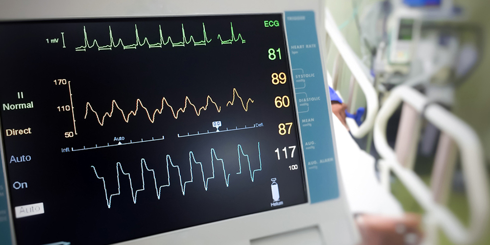
ECG
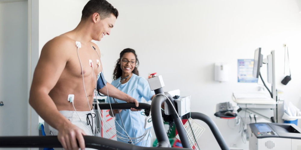
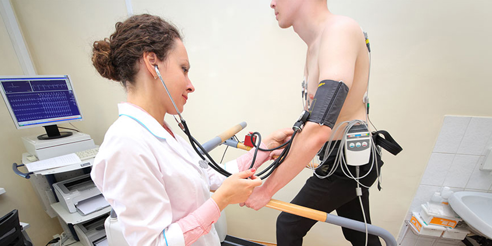
An electrocardiogram is a painless, noninvasive test to diagnose many common heart issues in human beings of all ages. Your doctor may also use an electrocardiogram to decide or locate: Abnormal rythm of heart (arrhythmias)
If blocked or narrowed arteries of your heart (coronary artery disorder) are inflicting angina (chest pain) or a heart attack
Nemo ipsam egestas volute and turpis dolores quaerat massa suscipit, luctus neque
Magna massa suscipit, luctus neque purus and ipsum neque dolor primis luctus tempor
An enim nullam tempor at pretium blandit
Magna massa suscipit, luctus neque purus and ipsum neque dolor primis luctus tempor
An enim nullam tempor at pretium blandit
Magna massa suscipit, luctus neque purus and ipsum neque dolor primis luctus tempor
P.ElectrocardiogramF.M
An electrocardiogram gives information about the electric signals of your heart. It's a an easy and painless proedure used to know about your heart's health.
Electrocardiogram — additionally known as ECGs or EKGs —
At Panchmegh Polyclinic & Diagnostic Centre, ECG test is carried out by a professional lady technician. At our center, reporting of the ECG is done by an Anaesthesiologist. Same day reporting and in a few urgent cases, instant reporting is also done..
Nemo ipsam egestas volute and turpis dolores quaerat massa suscipit, luctus neque
Magna massa suscipit, luctus neque purus and ipsum neque dolor primis luctus tempor
An enim nullam tempor at pretium blandit
Magna massa suscipit, luctus neque purus and ipsum neque dolor primis luctus tempor
An enim nullam tempor at pretium blandit
Magna massa suscipit, luctus neque purus and ipsum neque dolor primis luctus tempor
Blood Test
Pathology means “the study of disease," and pathologists are the doctors who interpret monitor laboratory testing, and help interpret those laboratory tests. At Panchmegh Polyclinic & Diagnostic Centre, the Department of Pathology and Laboratory Medicine is divided into two sections: Haematology and Biochemistry.Clinical Pathology is responsible for tests performed from blood, urine, sputum, semen and other body fluids.
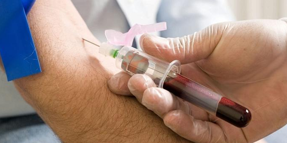
We perform blood tests under external quality control of CMC Vellore. We run control samples daily and croos check it with the results. Being a Doctor owned clinic, quality reporting is our primary motto. we understand the value of a diagnosis, on which treatment of a patients rely. Not a single report is delivered to the patients without a cross check by our pathologists.
Almost all types of blood tests are being carried out here. We do most tests at our lab
However, few tests are beyond our scope which we need to outsource to a higher centre
Instant delivery of reports is possible in case of emergency
For outsouced tests, delivery of reports are usually done within 48 hrs.
We perform blood tests under external quality control of CMC Vellore.
Clinical Pathology is responsible for tests performed from blood, urine, sputum, semen and other body fluids.